2016年9月6日火曜日

Is it possible to know the change of epidermis with the naked eye?


Topical steroids can cause atrophy of the skin as a side effect. The skin consists of epidermis and dermis. The epidermis as an outer layer is about 0.2mm in thickness which is very thin.  The atrophy of the dermis can be identified as roughness to a certain extent with the naked eye. However, can one identify the atrophy of epidermis with the naked eye?

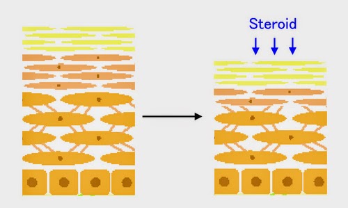
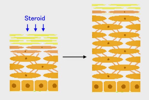
Before discussing the matter, I describe the histological structure of the epidermis. What provides the thickness of epidermis is Stratum spinosum which consists of 5-10 cell layers. When the number of cell layers decreases, the epidermis becomes atrophic. When it increases, the epidermis becomes hypertrophic. Above it exists Granular layer which consists of 2-3 cell layers. When it decreases or disappears, or when the blue-stained nuclei remain in the Stratum corneum over the Granular layer (parakeratosis), the skin is insufficiently keratinized and functionally defective.


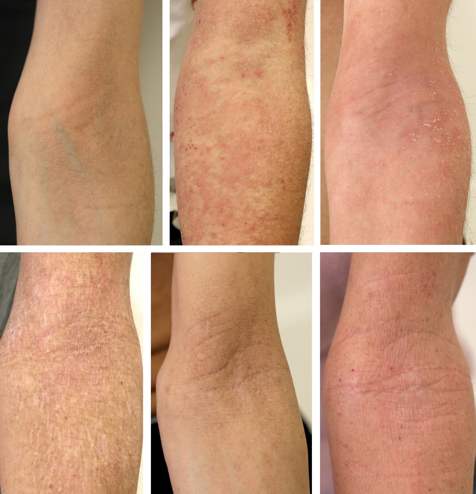
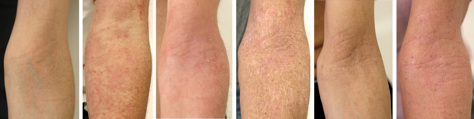
Here is a question. The following six photos of the elbows of different patients contain two atrophic epidermis and two severe insufficient keratinization. Can you identify which they are?


The following is the answer. Stop proceeding to read for a while if you like to think by yourself first.


☆ーーーー☆ーーーー☆ーーーー☆ーーーー☆ーーーー☆


The following is HE stain (left) and anti-cortisol antibody stain (right) of the six patients.


The first and the second patients are topical steroid users. The other four patients have withdrawn from topical steroids. The atrophy of epidermis can be seen in the first and fifth cases and the hypertrophy of epidermis in the third, fourth and sixth cases. Severe insufficiency of keratinization can be seen in the third and fourth cases.

Though the patients are different, the photos are arranged according to the stage of the disease. The first patient is using topical steroids. Though the appearance of the skin is normal, the epidermis is atrophic histologically. The second patient is just unable to control by topical steroids and anti-cortisol staining of the epidermis revealed that there are obvious patty defects. The third and fourth are in the situation that hypertrophy and insufficient keratinization of the epidermis are prominent. They are on rebound which has been prolonged.  The fifth has withdrawn from topical steroids but is easy to develop eczema even by a subtle stimulation as an aftermath or because of the natively thin skin. The sixth is normal in keratinization though the epidermis is hypertrophic. It is important to seek for any aggravating factor because allergic reaction to something might exist for a long time.
The clinical appearance looks like the process of withdrawal from topical steroids by arranging the photos from the left to the right though the photos are of different patients.


It is right that the epidermis is thin and its atrophy can’t be identified by the naked eye. However, it might be possible to discriminate to a certain extent by the disease phase in fact.

By the way, there are materials which regulate the thickness and differentiation of the epidermis. They are hyaluronic acid with intermediary molecular weight of around 100K Da and that with high molecular weight of over one million Da.


(Bourguignon et al. J Dermatol Sci. 2013 Oct;72(1):32-44. https://www.ncbi.nlm.nih.gov/pubmed/23790635)

Corticosteroids (cortisol) also regulate the thickness and differentiation of the epidermis. Moreover, aging also makes the keratinocyte atrophic.

   Three factors to regulate the thickness and differentiation of the epidermis

   ・Hyaluronic acid ( produced by keratinocyte itself, or topically applied)

   ・Cortisol ( produced by keratinocyte itself, or topically applied)

   ・Aging

These three factors are intertwined. Though aging decreases the production of cortisol in keratinocytes, the thickness of the epidermis never increases by it and rather decreases. The atrophy of the epidermis due to topical steroids is competed with hyaluronic acid with intermediary molecular weight.
Therefore it must be helpful to apply intermediary-sized hyaluronic acid to epidermal atrophy and high molecular weight hyaluronic acid to insufficient keratinization even if hyaluronic acid itself has no ability to induce cortisol production by keratinocytes.

As there was no available lotion containing hyaluronic acid with around 100k Da which is an intermediary molecular weight in Japan, I commercialized it as a cosmetics naming “Hyaluprotect”.


Though there are many available cosmetics containing hyaluronic acid with high molecular weight in Japan, there is no product disclosing its density. As hyaluronic acid is rather an expensive material, most of them compensate the viscosity by glycerol.
So I tentatively commercialized 1% aqueous solution of hyaluronic acid with a high molecular weight (two million Da) naming as “Hyaluprotect 200”. As there were considerable patients who preferred Hyaluprotect 200, I decided to manufacture a large bottle containing 150 ml.


The most rational way to select the lotion is confirming the status of epidermis by skin biopsy. In cases of above six patients, the first and the fifth cases with atrophic epidermis are suited to Hyaluprotect while the third and the fourth with prominent insufficiency of keratinization are suited to Hyaluprotect 200.

I will offer five 30ml bottles at the same price as the 150 ml bottle until the large bottles are available. For new customers, I recommend trying one by one of Hyaluprotect and Hyaluprotect 200 referring to the above photos (Remember that they are only a suggestion.)  You can use both at the same time.

(2016/09/06)

Sorry, the comment column is not available now. But the author believes readers can find some suggestion to overcome their own situations by the previous comments. 

2016年7月5日火曜日

My recent articles


I am sorry I have closed my comment column for a long time. It is rather a hard work for me to contact with real patients even at present because it recalls my old days when I saw patients with TSA day after day though my physical and mental status was the worst. As I experienced flashbacks, I am now closing the comment column.
However, I can investigate TSA or atopic dermatitis with the help of my friendly doctors. Or I can write some articles by remembering my own old experiences. Those works seem not to damage my health maybe because they are generalized ones. The followings are such works. I believe they are of some help for patients who are confused and lonely now.

"A prospective study of atopic dermatitis managed without topical corticosteroids for a 6-month period. " Click the following URL and you can read the whole content free of charge
https://www.dovepress.com/articles.php?article_id=27740
I believe it is an epoch-making study in which we followed 300 patients with atopic dermatitis without use of topical steroids.
You can watch an abstract movie also.
https://www.youtube.com/watch?v=TmDK3dfxNu4

And here is a movie titled “Concise guidelines for the management of atopic dermatitis”. This is a summarized movie about how guidelines for atopic dermatitis should be from the viewpoint of prevention and overcoming TSA.
https://www.youtube.com/watch?v=cpLosjKbsAI

The following one is about the mechanism of TSA. As the sample size of the study is very small, I admit it is still a hypothesis. But it is consistent with various phenomena about TSA and therefore I believe it contains some truth.
“Histological and Immunohistological Findings Using Anti-Cortisol Antibody in Atopic Dermatitis with Topical Steroid Addiction”
http://www.ncbi.nlm.nih.gov/pmc/articles/PMC4799038/
(2016/07/05 )

Sorry, the comment column is not available now. But the author believes readers can find some hints to overcome their own situations by the previous comments.

2014年11月10日月曜日

Eczema of infants may be prevented by moisturizing while moisturizing might disturb improvement of eczema

An article was published in the wall street journal this Nov. The title is “Are you bathing your baby too much?”
You can read the whole contents in the following site.

http://online.wsj.com/articles/are-you-bathing-your-baby-too-much-1415031555

In the article, a recent paper of Dr. Simpson is introduced. It revealed that high-risk infants of eczema were prevented from developing illness by frequent application of emollient.
http://www.jacionline.org/article/S0091-6749(14)01118-X/abstract

On the other hand, some patients with eczema, especially TSA/W patients, improve by stopping emollients as I have already written.
http://mototsugufukaya.blogspot.jp/2013/06/is-moisturizing-really-help-to-cure.html

It sounds controversy or very confusing. But it is not so difficult to explain logically. It only means that avoidance of the risk of oncet is different from the way how to treat the illness.

I explain it to be concrete. If I had two babies and one was with eczema while the other not, I would use emollient frequently for the purpose of prevention of eczema to the latter. But I might not use emollient to the baby with eczema because I consider maturartion of the skin is disturbed by it.
Emollient is supposed to prevent immunological sensitization of various antigens in the immature skin of high risk babies. On the other hand, emollient may delay maturation of skin against environmental dryness. In eczema-undeveloped babies, the merit of emollient is greater than demerit while in eczema-developed babies demerit might exceed merit because they have already been sensitized.

I don’t recommend using too much soaps or detergents to both babies. It is because they remove natural emollient which is produced by babies themselves. On the other hand, I recommend adequate disinfectant therapy in patients with eczema.
http://mototsugufukaya.blogspot.jp/2013/06/disinfectant-therapy-to-staphs-on.html
Soaps are different from disinfectants. I am afraid that might be another confusing problem among patients attention should be called to.
 
Sorry, the comment column is not available now. But the author believes readers can find some hints to overcome their own situations by the previous comments.

2014年7月16日水曜日

Topical steroids cause two kinds of atrophy – the epidermal atrophy and the dermal atrophy

  It is well known that topical steroids cause atrophy of the skin. However few people recognize that there are two kinds of atrophy. They are the epidermal atrophy and the dermal atrophy. 

The skin consists of three layers, i.e., epidermis, dermis and subcutaneous tissue. Topical steroids affect epidermis and dermis. The turnover of the epidermis is about 4-8 weeks while the dermis much longer. So the effect of topical steroids appears on the epidermis first. The epidermis becomes atrophic after two weeks’ application of topical steroids as shown in the following photo.

 Upper: before application of topical steroids. Lower: after application of topical steroids for two weeks.

 The macroscopic appearance of the epidermal atrophy can’t be distinguished from the normal skin. The thickness of the normal epidermis is about 0.2mm which is equal to the thickness of two sheets of copy paper. The thickness of the atrophic epidermis is equal to that of one copy paper. One can’t discriminate the difference of the two only by observation from outside though functional barrier ability is weakened in the atrophic epidermis of course.

 So the atrophic skin which is to be imaged by patients is not the epidermal atrophy but the dermal atrophy. The appearance of the dermal atrophy is so called paper-like skin, fine wrinkles or unevenness of the skin between follicles.
 Macroscopic appearence of the dermal atrophy

 The dermal atrophy is developed by the decrease of collagens in the dermis. The collagens are produced by fibroblasts very slowly and steroids suppress the activity of the production. So the dermal atrophy becomes visible after long-term application of steroids for more than several months or years. There is no mechanism which causes the dermal atrophy in atopic dermatitis itself. So I suspect most of dermal atrophy is the result of long term use of the topical steroids.

 The illustrated conceptual explanation of the two kinds of atrophy is as follows.

 When we discuss the atrophy of the skin due to topical steroids, we should be careful which of the atrophy is discussed. For example, one person might say ‘I don’t believe the skin atrophy occurs in only two weeks.’  He must be talking about the atrophy of the dermis. He is right in the dermal atrophy but the epidermal atrophy surely occurs in every human being in only two weeks though it is not visible and instinctively unacceptable.

 The epidermal atrophy occurs more rapidly than the dermal atrophy and it is reversibly recovered by discontinuance of topical steroids. The dermal atrophy is hard to be developed but the recovery after withdrawal from steroids is also slow and delayed. Both atrophies occur by aging also. Maybe the mechanism of atrophy due to steroids and aging is alike.

Hyaluronan (HA) lotion works on keratinocytes through the receptor of CD44 against atrophy due to steroids. How can the atrophy of the dermis be treated other than the passing of time? There is one method which I know, i.e., the PRP (platelet rich plasma) therapy.

PRP is rather common in the field of sports medicine or anti-aging medicine. It is rather an expensive treatment. However, I developed the economic method for preparing PRP. Please refer to the medical article which I published last month .
 http://journals.lww.com/prsgo/Citation/2014/06000/A_New_Economic_Method_for_Preparing_Platelet_rich.7.aspx

Platelets contain several growth factors which stimulate fibroblasts in the dermis. The activated fibroblasts produce collagens for several months and the macroscopic appearance is improved.
The following photos are before and after the treatment.

  Before PRP treatment

 Two months after PRP treatment

  The enlarged before and after photos.

 I believe the PRP therapy is very safe because PRP is prepared from the blood of the patient one’s self. I also believe the cost of PRP therapy becomes much more reasonable by adopting my method.
 There is a Youtube movie which I uploaded showing how PRP can be prepared by my economic method.
https://www.youtube.com/watch?v=gpeikkJhm0s

 The mainstay for the healing of topical steroid addiction is the time. It is surely the safest one but there are some other useful methods. PRP therapy is one of them for patients annoying delayed recovery of the dermal atrophy.
 Time can heal the topical steroid addiction. But time is not only one which helps your recovery. Believe and try to find your own way to exit.

==========
P.S. A figure from Dr. Sato’s medical article written in 1996 (As an answer to Dan.D in the below comment)

2014年6月6日金曜日

The skin produces steroids by itself

 It has been revealed that the skin (keratinocytes) itself has an ability of producing steroids. It is a proved fact.

The skin becomes thin by continuous application of steroids, temporarily hypertrophic (thicker than normal) after withdrawal and normalized after a significant period.
 The consecutive changes can be explained as follows from the thinking of cortisol production by the skin. It is not proved yet. It is my hypothesis but it can explain various phenomena about TSA.

 Topical steroids make the epidermis thin by suppressing the proliferation and differentiation of the keratinocytes.

 Physiologically, the epidermis produces steroids. The produced (inner) steroids regulate the proliferation and differentiation of the skin. However, prolonged application of topical steroids may suppress the production of the inner steroids. It is just like adrenal grand is suppressed by systemic steroids administration.

 When topical steroids were withdrawn and the inner steroids can’t be produced immediately, the epidermis becomes thickened and poorly differentiated. At that period skin can’t work as a normal organ and may react to various stimuli.

 After a significant time has passed, the skin recovers its function to produce inner steroids. That is the story of TSW from the viewpoint of steroid production by the skin itself.


From the above theory, many phenomena can be explained in an integrated way.

For example, ultraviolet rays are proved to induce the production of steroids in the skin. It is empirically known that UV therapy works on eczema and it induces no rebound. It may be because UV never suppresses inner production of steroids but increases it.

 The so called “moisturizer withdrawal therapy” is another example. The epidermis is proved to produce more cortisol in the dry atmosphere by the experiment using cultured keratinocytes(→ ref.). When the procedure of TSW prolongs and the redness of the skin continues, withdrawal from the whole topical agents sometimes works though it is accompanied by another rebound just like withdrawal from steroids. The mechanism may be the facilitation of recovery of production of inner steroids.

 Cholesterol is the primary material of all steroids. It is converted by the enzyme CYP11A1 to pregnenolone. On the other hand, CPY11A1 is an enzyme to metabolize aromatic carbonates which are contained in Coal Tar. Coal Tar has been known to be useful to make eczema subside since the time before topical steroids were on the market. If aromatic carbonates in the coal tar induce expression of CPY11A1, cortisol production in the skin is also accelerated. Vitamin D is also known to be metabolized by CPY11A1 (→ ref.) and there is medical literature topical Vitamin D works on eczema (→ ref.) . It may be of the same reason.

 Nowadays it is revealed that there are three organs which produce steroids, i.e. adrenal grand, brain and skin. The fact was elucidated only several years ago and it might not be well known to the ordinary people. The skin also produces sex hormone also. That is why women’s skin changes according to their periods.

 There are patients who used topical steroids so long a time and the epidermis seems not to produce any more steroids eternally.  I believe there are no patients who can’t recover the inner production of cortisol within the skin but you can choose the method of supplying steroids as much as needed by topical agents.

 It is so much for today. I hope your itching disappeared or decreased for a little time by reading my article
 I know you sufferers through TSW are spending a severe time day by day. But there is an exit because your skin is living and struggling to reconstruct the balance for recovery.

PS: Suppliment for Brenda Chu.
Cortisol synthesis pathway is in the following figure.
Cortisol is made from cholesterol via pregnenolone by the enzymes of CYP11A1, CYP17, 3βHSD, CYP21 and CYP11B1. Progesterone is on another pathway and doesn’t convert to cortisol.
I suppose any agent which induces expression of CYP11A1, CYP17, 3βHSD, CYP21 or CYP11B1 can be a facilitator of TSW. The examples are coal tar and topical Vitamin D above mentioned.


Please click here as for the clofibrate ointment. It is a non-steroidal agent with mild anti-inflammatory effect.

2014年5月28日水曜日

The rejuvenating effect of the aged skin by Dr. Fukaya’s skin repair lotion was published as a medical paper.

 One year has passed since Dr. Fukaya’s skin repair lotion went on the market. The main component of the lotion is around 100k Dalton-sized hyaluronan which has been confirmed to rejuvenate the aged skin. As the data is of the component and not of the lotion itself, the lotion was studied if it really rejuvenates the aged skin.
http://www.scirp.org/journal/PaperInformation.aspx?PaperID=46333 
 (It is an open access journal and everyone can download the whole contents. Click the above figure of the journal and move to the download site.)

 The following is the summarized content. Sorry for a little difficulty in understanding due to specialty.
 10 healthy aged individuals over 65 years old were enrolled. They were indicated to apply the lotion twice a day for 14 consecutive days on the neck. The skin was biopsied before and after the 14 days’ application and immmuno-histologically examined.

 The results were as the table.

  PCNA(proliferative cell nuclear antigen) is a marker of cell division and proliferation. Filaggrin is the main component protein of the corneal layer. 11βHSD1 and 2 are a little complicated to be explained. It has been revealed that the skin itself produces steroids (cortisol). The 11βHSD1 is associated with the activation of steroids while 11βHSD2 is with its inactivation.  If 11βHSD1 decreases or 11βHSD2 increases after application of the hyaluronan lotion, it means that steroids within the epidermis is suppressed by the lotion and that can be one reason why the hyaluronan lotion make the atrophic skin recover.

 The results show that PCNA increased in 8/10 cases, filaggrin increased in 6/10, 11βHSD1 decreased in 5/10 and  11βHSD2 increased in 5/10. The rate of the cases in which  11βHSD1 decreased or 11βHSD2 increased was 8/10.
The real images of immunohistochemical staining are as follows  in case 1 for example.

 a: PCNA, b: filaggrin, c: 11βHSD1,d: 11βHSD2. Upper: before application, lower: after application. The brown color means positive findings.

 From the above experiments, it was concluded that 1) Dr. Fukaya’s skin repair lotion has an efficacy of rejuvenating the aged skin. 2) The mechanism might be shifting 11βHSD1/2 balance.

 Though the above study is about the aged skin of healthy individuals, it can be interpreted to the steroid-induced atrophy. It is because the atrophic skin due to steroids is also reported to recover by the same Dalton sized hyaluronan as the aged skin.

 By the way, considering the fact that the aged skin and the atrophic skin due to steroids can both recover by the same hyaluronan, I suggest that the following can be said.

To apply topical steroids to the skin is very near to making the skin age.

 The difference is that aging can’t be removed while topical steroids can be withdrawn. Anyway, I strogly recommend my hyaluronan lotion to all steroid-users. It might be a little expensive but it really works to prevent atrophy.

 Don’t misunderstand me that I am only a merchant of my lotion. I am enough satisfied with my present successful situation and income as a cosmetic surgeon. The reason why I am transmitting information about TSA or suggesting my lotion is my outside interests.
Moreover, as a dermatologist also, it is really exciting to know the mechanism of the skin by which topical steroid addiction might be caused.
 
☆===☆=== ☆===☆===☆===☆
 The folowings are addition in response to the comment of Javeriya Iftekhar.
 Patients after long TSW period present more aged appearance than the real age because the swelling enlarges the skin especially around the eyelids.
 Of course my lotion doesn’t improve this kind of sagging. The excessed skin should be excised surgically in such cases.

Just affter the upper eyelids surgery.


One week after the upper surgery and just after the lower eyelids surgery.

 
Two weeks after the upper surgery and one week after the lower eyelids surgery.


 The price in my clinic is JPN 120,000 (about US$ 1,200) for the upper eyelids and JPN180,000 (about US$ 1,800) for the lower eyelids. I think they are absolutely reasonable. That is my everyday work. I don’t see patients with TSW or AD because I don’t like to charge money for only an advice. However, you are welcome if you like to make your appearance younger surgically.
 This kind of work really keeps my mind healthy because I can charge money for my real skills and the result appears in only one week. It is too hard for doctors also to wait until the natural recovery comes with very few medical intervention. That is why I changed my work. But I am still transmitting information through internet like this because I know very well that my knowledge about TSW is precious for patients.

 There is another option for improving finer lines. I will address it next time.
 
  Please click here as for the clofibrate ointment. It is a non-steroidal agent with mild anti-inflammatory effect.

 Sorry, the comment column is not available now. But the author believes readers can find some hints to overcome their own situations by the previous comments.

2014年5月19日月曜日

AAD (American Academy of Dermatology) guideline of atopic dermatitis has been revised.

 There is unfortunate news. In the new guideline published in May 2014, Topical Steroid Addiction(TSA) or Red Burning Skin Syndrome(RBSS) was not referred to as a side effect of topical corticosteroids(TCS).
 My former article about the significance of AAD guideline is in the following URL.
http://mototsugufukaya.blogspot.jp/2013/06/aad-american-academy-of-dermatology.html
 As the sunset of the new guideline is 2019 and the interval period since the sunset of the last guideline was 5 years, we must struggle and prepare for another 10 years to add TSA or RBSS to the next guideline. It must be really a hard work but we just have to do it.
In the new guideline, the section about topical corticosteroids is described as the followings (brown,italic). You can read or download the full article in the AAD site.
http://www.aad.org/education/clinical-guidelines

 I will add my comment after each paragraph.

TOPICAL CORTICOSTEROIDS
 TCS are used in the management of AD in both adults and children and are the mainstay of anti-inflammatory therapy. They act on a variety of immune cells, including T lymphocytes, monocytes, macrophages, and dendritic cells, interfering with antigen processing and suppressing the release of proinflammatory cytokines. They are typically introduced into the treatment regimen after failure of lesions to respond to good skin care and regular use of moisturizers alone.


 The last phase is important from the viewpoint of prevention of TSA. I agree in that TCS should not be regarded as the first choice.

Efficacy
 TCS have been used to treat AD for more than 60 years. Their efficacy has been demonstrated with a wide variety of preparations and strengths, with more than 110 different RCTs performed to date. They are generally the standard to which other topical anti-inflammatory therapies are compared. In addition to decreasing acute and chronic signs of AD, multiple trials have shown decreased pruritus with their application. TCS are used for both active inflammatory disease and for prevention of relapses. Comparative trials are limited in duration and scope (ie, they mainly involve, and occasionally, agents), and as a result, there are no data to support or a few specific agents as being more efficacious than others. Patient vehicle preference, along with cost and availability, often determine their selection. A summary of recommendations on TCS use is in Table IV, with the level of evidence in Table III.


Dosage
 TCS are grouped into 7 classes, from very low/lowest potency (VII) to very high potency (I), based on vasoconstriction assays. Table V provides some representative examples of available agents in each class. There is a paucity of studies that examine a range of TCS doses in large numbers of patients and with the lack of an established optimum, great variability in dosing exists. Some use a short burst of a high-potency TCS to rapidly control active disease, followed by a quick taper in potency, whereas others use the lowest-potency agent thought to be needed and adjust upward only if this fails.
 No universal standard exists for quantity of application, although suggested methods include use of the adult fingertip unit (the amount from the distal interphalangeal joint to the fingertip, or approximately 0.5 g, being applied over an area equal to 2 adult palms), following the rule of 9’s that measures the percent affected area, and use of charts that propose amounts based on patient age and body site.
Children have a proportionately greater body surface area to weight ratio, and as a result, have a higher degree of absorption for the same amount applied. But during significant acute flares, the use of mid- or higher-potency TCS for short courses may be appropriate to gain rapid control of symptoms, even in children.  However, for long-term management, the least-potent corticosteroid that is effective should be used to minimize the risk of adverse effects. Greater caution regarding TCS potency is also needed when treating thin skin sites (ie, face, neck, and other skin folds), where there is greater penetration and higher likelihood for systemic absorption. It is important to monitor quantities of TCS used over time, which may impact efficacy and safety.


 There is a little conflicting expression in the above paragraph. It says significant acute flares should be suppressed by potent TCS while they should be limited to be used for long-term management. The phase sounds beautifully harmonized, but is it really feasible in fact?

Frequency of application
 Most studies on the efficacy of TCS in AD management involve twice-daily application. This is the most common clinical practice and also the generally recommended frequency. However, there is evidence to suggest that once-daily application of some potent corticosteroids may be as effective as twice-daily application. Some newer formulations also use once-daily application. For acute flares, use of TCS is recommended every day until the inflammatory lesions are significantly improved and less thick, for up to several weeks at a time. After obtaining control of an outbreak, the goal is to prolong the period until the next flare.
Previously, TCS use was stopped on improvement of symptoms and signs of disease, switching to the use of moisturizers alone and reinstituting the TCS only with subsequent relapses. However, in recent years, a more proactive approach to maintenance has been advocated for those patients who experience frequent, repeated outbreaks at the same body sites. This entails the scheduled application of a TCS once to twice weekly at these particular locations, a method that has reduced rates of relapse and increased time to first flare relative to the use of moisturizers alone (to be discussed further in part 4 of these guidelines).


 The so-called proactive approach is described here. It is a method that was studied from the viewpoint of health economics. In the study patients were to be controlled by potent TCS before entry of the study. Successfully controlled patients were divided into two groups, that is, patients who use TCS regularly 1-2 days per week (proactive approach) and the others using TCS only when the flare was developed (reactive approach). In the former the interval between relapses was longer and economic burden was less.
 However, there are two points to be noted in translating the study into clinical practice. 1) The enrolled patients in the study were all successfully controlled ones by the initial treatment by potent TCS. It means patients who were judged as poorly controlled at the initial stage were not enrolled. 2) According to the proactive approach, patients can’t stop TCS theoretically forever. Eczema sufferers often become healed naturally especially in children. But in the approach such natural healing is not assumed. 

Adverse effects and monitoring
 The incidence of reported side effects from TCS use is low; however, most studies fail to follow up patients long term for potential complications. Cutaneous side effects include purpura, telangiectasia, striae, focal hypertrichosis, and acneform or rosacea-like eruptions. Of greatest concern is skin atrophy, which can be induced by any TCS, though higher-potency agents, occlusion, use on thinner skin, and older patient age increase this risk. Many of these side effects will resolve after discontinuing TCS use, but may take months. Sites of treatment should be assessed regularly for these adverse effects, particularly with use of more potent agents. Continuous application of TCS for long periods of time should be avoided, to limit the occurrence of negative changes. Proactive, once to twice weekly application of mid-potency TCS for up to 40 weeks has not demonstrated these adverse events in clinical trials.


TCS application on AD lesions does reduce Staphylococcus aureus bacterial load, likely via decreasing the inflammatory cytokines that inhibit antimicrobial peptide production. There is some worry that TCS may impair the process of wound healing and re-epithelialization, although excoriated and fissured lesions should be included in treatment given that the underlying inflammation and pruritus lead to these secondary changes. Allergic contact dermatitis/type IV hypersensitivity can develop to TCS or other ingredients in their formulations, such as propylene glycol and preservatives. This should be considered if lesions fail to respond as expected or worsen with application. Patch testing is needed to determine if the allergen is the steroid compound itself or a component of the vehicle. Development of tachyphylaxis is of concern for some practitioners, where the efficacy is thought to decrease with repeated use of the same agent, although data are lacking to suggest that this is a significant clinical problem. Although there is documented risk with systemic corticosteroid use, an association between topical steroid use and the development of cataracts or glaucoma is not as clear. Nonetheless, minimizing use at periocular sites may be prudent.

 Although the term “tachyphylaxis” is not appropriate to represent TSA or RBSS, the above phrase might refer to it. (Please refer to my article in the following URL about the difference of both.
http://topicalsteroidaddiction.weebly.com/chapter-1512288tragedy-of-tachyphylaxis.html)
 If the “ tachyphylaxis” here is used in the original meaning as epinephrine becomes weakened to the blood vessels in efficacy, the above phrase is true. Tachyphylaxis of TCS must be of little importance in clinical practice. But if the authors use the term as the representative of TSA or RBSS, the phrase “data are lacking to suggest that this is a significant clinical problem” is wrong. There are several medical case reports for examples by me or Dr Rapaport.
If the authors confuse tachyphylaxis and TSA or RBSS, it expresses ironically the fact that most dermatologists have not experienced to see patients at withdrawal from TCS. It is because tachyphylaxis resembles the situation before stopping TCS while TSA or RBSS is the term expressing the rebound after withdrawal.
 In the world of medical journals or in the practicing rooms of dermatologists, there might be very few TSA sufferers. That is probably because they don’t visit dermatologists. But is it really “data are lacking to suggest that this is a significant clinical problem”?  Is the number or comments of ITSAN’s member not a data for example?

 Topically applied corticosteroids, particularly high- and very high potency agents, can be absorbed at a degree sufficient to cause systemic side effects. The risk of hypothalamic-pituitary-adrenal axis suppression is low but increases with prolonged continuous use, especially in individuals receiving corticosteroids concurrently in other forms (inhaled, intranasal, or oral). As discussed above, children are more susceptible as a result of a greater body surface to weight ratio. There is also some concern for negative effects on linear growth, although reports have given mixed conclusions. Hyperglycemia and hypertension have rarely been reported.
 A systematic review concluded that TCS overall have a good safety profile. No specific monitoring for systemic side effects is recommended for patients with AD at this time. However, if hypothalamic-pituitary-adrenal axis suppression is a concern, this can be assessed by performing a cortisol stimulation test to check for appropriate adrenal response. As discussed in part 1 of these guidelines, some children with AD are underweight as a result of severe disease, although further decline in growth should prompt consideration for investigation.


Addressing concerns with TCS use
 Although judicious use of TCS is certainly warranted, recognition of undertreatment as a result of steroid phobia is also important. One survey of 200 dermatology outpatients with AD found that 72.5% were worried about use of TCS on their own or their child’s skin, with 24% admitting
noncompliance with therapy as a result of these concerns. Other studies have shown that patient knowledge of steroid class potencies is poor and leads to inappropriate use. Thus, to achieve good response, it is important to address such fears and incorrect beliefs. The risks associated with TCS use do appear to be low with appropriate application and choice of potency, combined with periods of nonuse. A higher strength of recommendation (than actual level of evidence) is therefore placed on counseling, because the benefits outweigh the risks.

 The undertreatment due to steroid phobia is taken up here. There exist such patients really. Some patients even believe their eczema must be healed only if they never use TCS. However, they might be true because atopic dermatitis has a tendency of self-healing originally and nobody can deny that TCS disturb it. Moreover, when the patient becomes addicted to TCS, the recovery can’t be obtained without the “undertreatment”.
 From the short term observation in the clinical practice, it certainly looks successful to treat the “undertreated” patients by TCS. That is why so many medical studies exist suggesting TCS are useful. However, is it successful even from the long-term viewpoint also? Hasn’t the number of patients with adulthood atopic dermatitis increased after the dermatologists began to prescribe TCS several decades ago?  Why do patients with atopic dermatitis only complain or worry about TCS use?  I have once researched about the problem. The patients’ phobia to TCS originated  mostly from their own experience
(http://topicalsteroidaddiction.weebly.com/chapter-1612288steroid-refusal-irrelevant-to-media-coverage.html). I suggest dermatologists should investigate the reason of the patients’ fear, not worrying about incompliance of the patients.

 Please click here as for the clofibrate ointment. It is a non-steroidal agent with mild anti-inflammatory effect.
 
Sorry, the comment column is not available now. But the author believes readers can find some hints to overcome their own situations by the previous comments.